

Intra-articular fractures of the calcaneus, particularly Sanders Type II through IV patterns, represent one of the most mechanically and biologically demanding injuries in orthopedic trauma. The calcaneus serves as the foundational heel strut of the human foot, transmitting immense axial loads while acting as the lever arm for the Achilles tendon. High-energy axial impact—most commonly from falls from height or motor vehicle accidents—drives the lateral process of the talus downward, splitting the posterior facet of the calcaneus, shattering the lateral wall, and collapsing the crucial Böhler's and Gissane angles. Historically, conservative management resulted in severe subtalar arthritis, broadened heel deformity, and catastrophic footwear impingement. Modern surgical intervention demands anatomical restoration of the subtalar joint surface and correction of heel height, width, and varus alignment. Today, trauma surgeons must navigate the critical decision between the classic extensile lateral "No-Touch" approach and the minimally invasive sinus tarsi approach, balancing the need for direct articular visualization against the grave risk of lateral soft tissue necrosis.
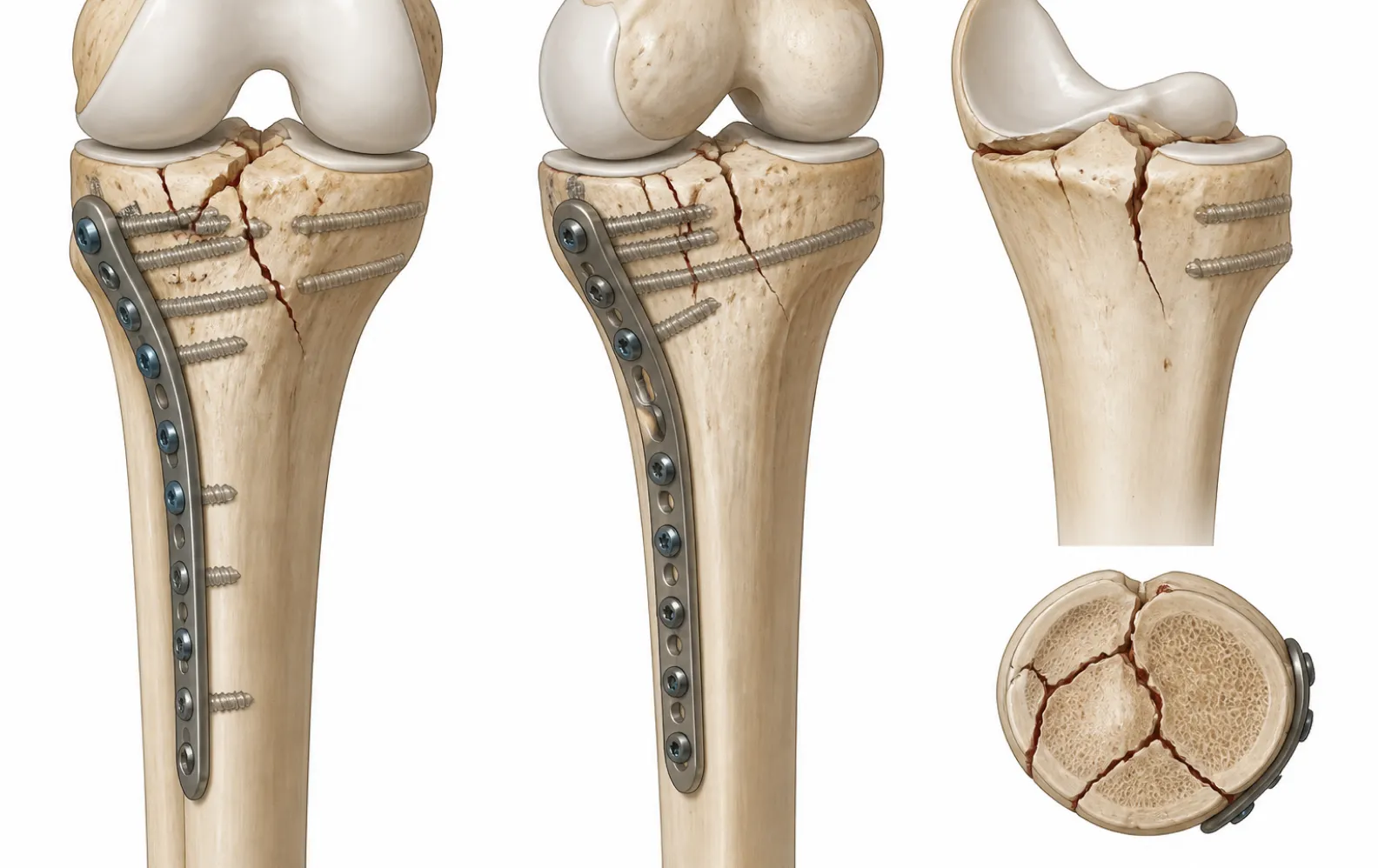
The Extensile Lateral L-Shaped Approach: The "No-Touch" Technique
The traditional gold standard for complex, highly comminuted calcaneal fractures (Sanders Type III and IV) is the extensile lateral L-shaped approach. This technique provides unparalleled, panoramic exposure of the entire lateral wall, the posterior facet, and the calcaneocuboid joint, enabling surgeons to reconstruct the shattered bone block under direct vision.
However, the lateral skin of the heel is uniquely vulnerable. Its blood supply relies on a delicate, fragile angiosome fed by the lateral calcaneal artery. Elevating this skin flap incorrectly invites catastrophic wound dehiscence and deep implant infection. To mitigate this, the "No-Touch" technique is mandatory.
The incision begins vertically just anterior to the Achilles tendon and curves sharply at a 90-degree angle just proximal to the glabrous (thickened) skin of the heel pad, extending anteriorly toward the base of the fifth metatarsal. The critical surgical maneuver requires taking the flap as a single, "full-thickness" osteocutaneous unit. The surgeon incises directly down to the periosteum of the lateral calcaneal wall. Rather than dissecting through subcutaneous fat, an elevator is used to peel the periosteum directly off the bone. Three to four 1.6mm Kirschner wires are driven into the lateral talus and bent backward, acting as self-retaining retractors to hold the thick flap up and out of the way. Throughout the procedure, the edges of the skin flap must never be touched, crushed with forceps, or subjected to prolonged manual retraction.
The Sinus Tarsi Approach: Minimally Invasive Direct Targeting
Because the extensile lateral approach carries wound complication rates historically reaching 15% to 25%, the sinus tarsi approach has gained tremendous traction for Sanders Type II and select Type III fractures. This minimally invasive technique sacrifices panoramic metaphyseal exposure in favor of preserving lateral vascular biology.
A straight 3cm to 5cm incision is made from the tip of the lateral malleolus extending anteriorly toward the fourth metatarsal base, directly overlying the sinus tarsi. This keyhole window provides a direct, uninhibited view of the crucial posterior facet—the most important articular surface in the subtalar joint. While the lateral wall blowout cannot be visually assembled piece by piece, surgeons use a combination of direct facet visualization and intraoperative fluoroscopy to achieve reduction.
The sinus tarsi approach drastically reduces postoperative wound necrosis and allows for earlier surgical intervention, often bypassing the typical 10-to-14-day "wrinkle sign" waiting period required for the swelling to subside before an extensile lateral incision can be safely executed.
Articular Reconstruction and the Schanz Pin Joystick Maneuver
Regardless of the approach, the sequence of reduction remains physically identical. The primary goal is to pull the depressed posterior facet out of the calcaneal body, restore the critical Böhler's angle (normally 20° to 40°), and recreate the anatomical Gissane angle.
To unlock the impacted fragments, a 4.0mm or 5.0mm Schanz pin is driven transversely into the dense bone of the posterior calcaneal tuberosity from the lateral side. Acting as a powerful joystick, the surgeon applies strong downward, posterior, and valgus traction on the Schanz pin. This maneuver pulls the tuberosity out of varus, restores heel height, and opens the internal void. With the tuberosity held in traction, an elevator is inserted beneath the depressed posterior facet. The facet is pushed upward until it sits perfectly flush with the opposing articular surface of the talus, confirmed by direct vision (or fluoroscopic Broden's views).
Once the articular block is reduced, it is pinned provisionally to the intact medial sustentacular fragment (the "constant" fragment) using K-wires. If a large metaphyseal void remains beneath the elevated posterior facet, structural bone graft, allograft chips, or synthetic bone substitutes must be packed into the defect to prevent late subsidence under weight-bearing loads.
Implant Selection: Conventional vs. Locking Calcaneal Plates
Once the calcaneus is reduced and pinned, an implant must bridge the tuberosity, the posterior facet, and the anterior process, compressing the lateral wall inward to restore normal heel width.
For the extensile lateral approach, a standard perimeter calcaneal plate is typically applied. These plates are manufactured from pliable, low-profile titanium or stainless steel, allowing the surgeon to manually bend and contour the struts to match the reconstructed lateral wall perfectly. In robust bone, non-locking cortical screws provide excellent interfragmentary compression, drawing the lateral wall medially.
However, in severely comminuted fractures or osteoporotic patients, conventional screws may pull out. In these scenarios, a calcaneal locking plate is deployed. Locking screws thread directly into the plate matrix, creating a fixed-angle construct that maintains the restored Böhler's angle without relying on frictional bone purchase. Modern locking plates often feature a hybrid design—utilizing locking screws to support the posterior facet and non-locking screws to compress the lateral wall.
For the sinus tarsi approach, standard perimeter plates cannot be inserted through the small keyhole incision. Instead, specialized low-profile locking plates, mini-fragment straight plates, or independent bone plate and screws (lag screws) are utilized. The surgeon slides the dedicated sinus tarsi plate submuscularly along the lateral wall, locking it in place percutaneously under fluoroscopic guidance to secure the articular reduction.
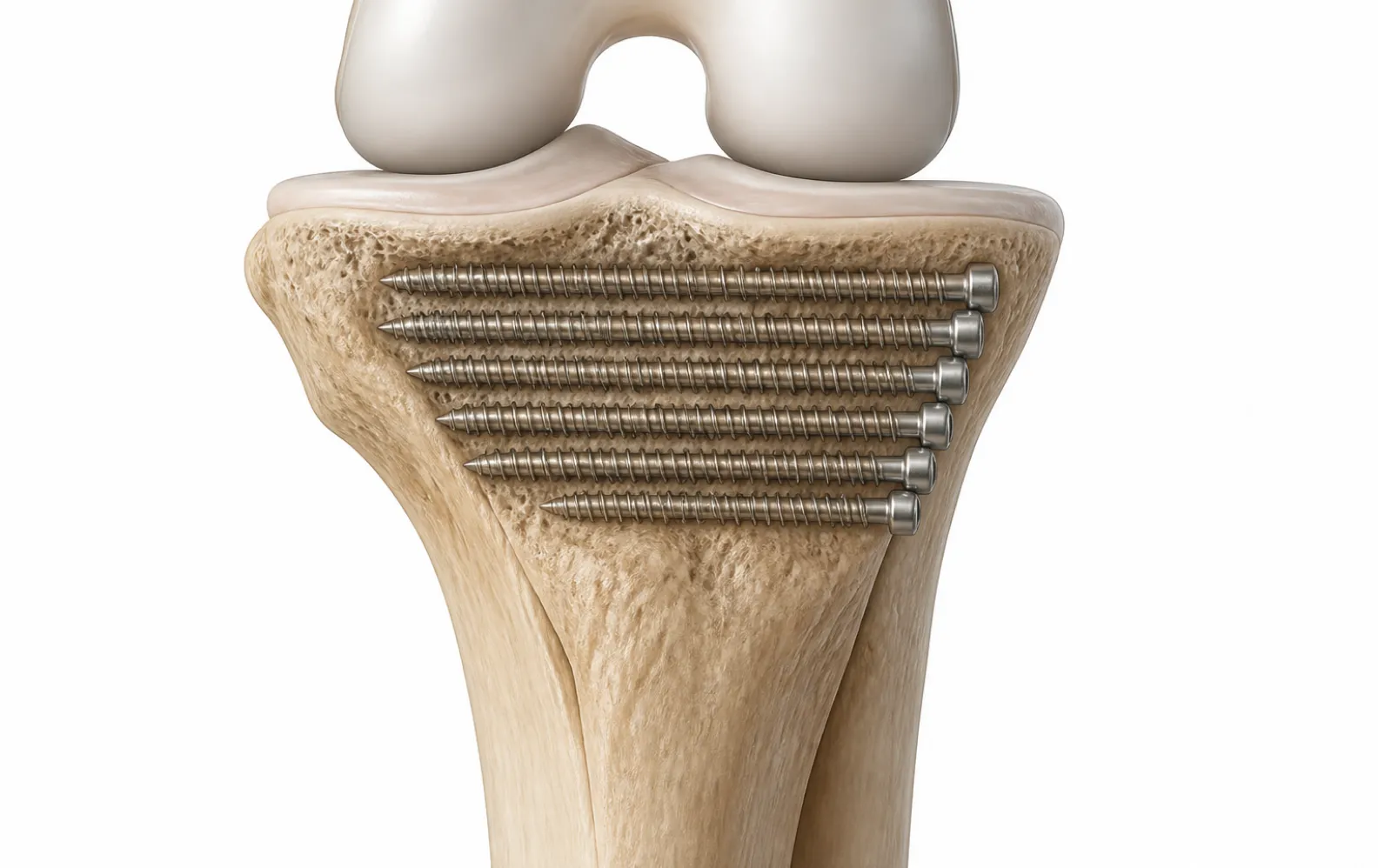
Biomechanical Trade-offs: Extensile vs. Sinus Tarsi
Selecting the optimal operative strategy requires evaluating both the fracture morphology and the patient's soft tissue envelope:
| Surgical Parameter | Extensile Lateral L-Shaped Approach | Minimally Invasive Sinus Tarsi Approach |
|---|---|---|
| Optimal Fracture Pattern | Sanders III & IV; Severe lateral wall blowouts | Sanders II & simple III; Joint depression types |
| Articular Exposure | Complete panoramic visualization of all facets | Excellent view of posterior facet; limited anterior view |
| Wound Complication Risk | High (10% - 25%); requires strict No-Touch technique | Low (<5%); preserves lateral angiosome |
| Timing of Surgery | Delayed 10–14 days (waiting for "wrinkle sign") | Early intervention possible (often <7 days) |
| Implant Matrix | Standard perimeter calcaneal locking/non-locking plates | Specialized sinus tarsi plates or independent lag screws |
Closure and Postoperative Protection
Wound closure in the extensile lateral approach is critical. The thick osteocutaneous flap is meticulously sutured using a modified Allgöwer-Donati technique. This specialized stitch passes through the dermal layer on the flap side without penetrating the fragile epidermis, drawing the viable skin edge tightly against the plantar boundary without strangulating capillary blood flow. A closed-suction drain is frequently placed deep to the flap to prevent hematoma accumulation, which could place immense pressure on the tenuous skin.
Postoperative rehabilitation emphasizes early, protected motion. The foot is placed in a bulky Jones splint in neutral position. To prevent catastrophic hardware pull-out and loss of Böhler's angle, strict non-weight-bearing must be enforced for 8 to 12 weeks, until radiographic consolidation of the posterior facet and tuberosity is confirmed.
Procurement Sourcing for Foot and Ankle Trauma
Because the clinical decision between an extensile approach and a sinus tarsi approach is frequently made dynamically based on soft tissue evolution, trauma centers must maintain comprehensive inventories. Sterile processing departments require immediate access to perimeter calcaneal plates in multiple lengths, sinus tarsi specific mini-plates, and complete 2.7mm/3.5mm foot and ankle screw matrices. Relying on loaner sets from original equipment manufacturers (OEMs) introduces unacceptable logistical delays when treating acute, severely swollen hindfoot trauma.
BoneCraft supports high-volume trauma facilities and global orthopedic distributors with immediate, ready-to-ship access to genuine, factory-sealed wholesale warehouse inventory. Our supply chain ensures that regional distributors and hospital purchasing networks maintain deep par levels of critical distal lateral fibula plate assemblies and complex hindfoot reconstruction systems. For complete access to verified, traceable Zimmer Biomet implants, contact our distribution team at Sale@bone-craft.com.
Frequently Asked Questions
What is the "No-Touch" technique in the extensile lateral approach?
The "No-Touch" technique is a mandatory soft-tissue management protocol designed to prevent catastrophic skin flap necrosis. Instead of using forceps or retractors on the skin edges, the surgeon develops a full-thickness osteocutaneous flap directly off the periosteum. K-wires are driven into the talus and bent backward to hold the flap open. The skin edges are never touched or manipulated throughout the duration of the surgery.
How is the Schanz pin joystick maneuver performed in calcaneus fractures?
A 4.0mm or 5.0mm Schanz pin is driven transversely from lateral to medial into the dense bone of the posterior calcaneal tuberosity. The surgeon uses this pin as a handle (joystick) to apply strong downward and posterior traction, pulling the tuberosity out of its varus, impacted position. This maneuver restores heel height, corrects varus deformity, and opens the internal bone void so the depressed posterior facet can be elevated.
Why has the sinus tarsi approach become increasingly popular for calcaneus fractures?
The sinus tarsi approach utilizes a small 3cm to 5cm incision directly over the subtalar joint. Unlike the extensile lateral approach, it preserves the fragile lateral calcaneal angiosome, dramatically lowering the risk of devastating wound dehiscence and deep infection (from up to 25% down to less than 5%). It provides excellent visualization of the critical posterior facet, though it limits complete panoramic exposure of the lateral wall.
What is the significance of restoring Böhler's angle during surgery?
Böhler's angle (normally 20° to 40°) measures the anatomical arch and height of the calcaneus on a lateral radiograph. In a fracture, the posterior facet collapses, flattening or reversing this angle. Restoring Böhler's angle is paramount; failure to do so leaves the heel shortened, causes the talus to sit horizontally, severely limits ankle dorsiflexion, and causes permanent mechanical impingement.
When is a calcaneal locking plate required instead of a conventional plate?
Conventional calcaneal plates rely on friction and non-locking screws, which are highly effective in dense, healthy bone. However, in severe comminution (highly shattered bone) or osteoporotic patients, non-locking screws cannot achieve sufficient purchase. A locking plate secures the screw heads directly into the plate body, creating a fixed-angle scaffold that physically props up the posterior facet and maintains Böhler's angle regardless of bone density.
Why is the Allgöwer-Donati suture technique used for lateral heel closure?
The lateral calcaneal skin flap has a highly precarious blood supply. Standard simple or mattress sutures can compress and strangulate the dermal capillaries, leading to edge necrosis. The modified Allgöwer-Donati suture is designed to pass through the dermis on the flap side without exiting the epidermis, providing secure closure tension while maximizing blood flow to the critical edge of the flap.
-

BoneCraft Medical & Technical Editorial Team
Orthopedic Fixation Specialists
The BoneCraft Editorial Team comprises biomedical specialists and medical device supply chain experts. Dedicated to surgical transparency and technical precision, we publish insights on trauma plating, intramedullary fixation, and biomechanical implant performance. Our content synthesizes established surgical techniques, clinical literature, and exact specifications for genuine Zimmer Biomet orthopedic products to support global healthcare distributors and procurement teams.
