

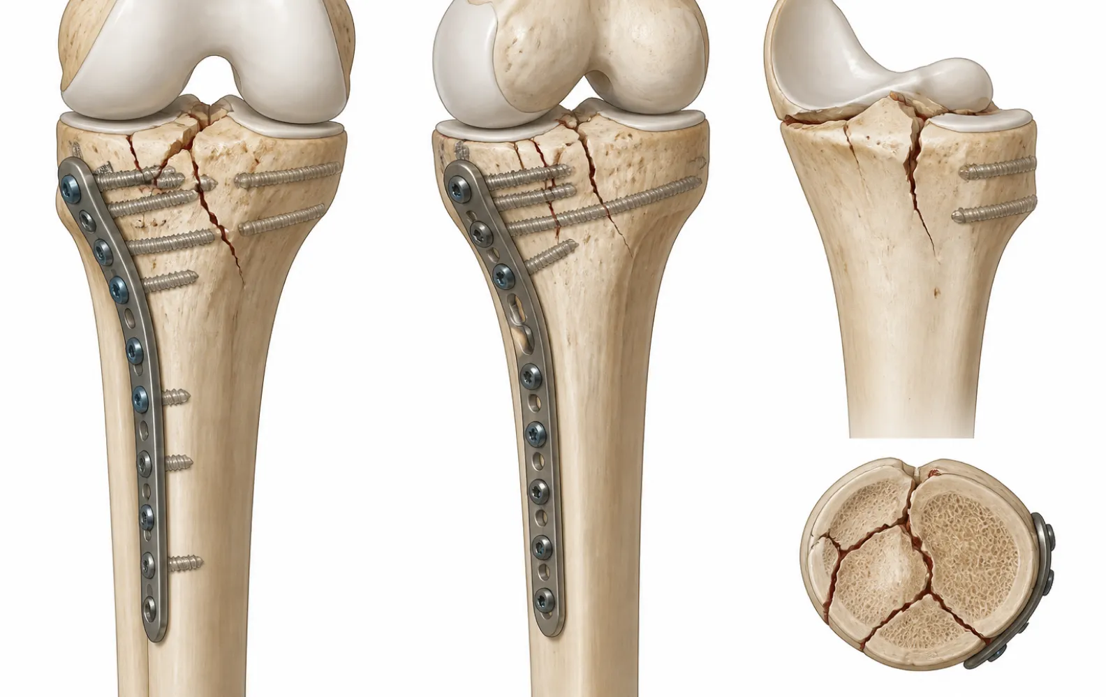
Fractures of the distal femur—encompassing AO/OTA 33-A extra-articular supracondylar fractures and AO/OTA 33-C complete intra-articular bicondylar fractures—represent severe mechanical disruptions of the lower extremity. Successful surgical management requires restoring the anatomical congruity of the knee joint, correcting coronal and sagittal axial alignment, and re-establishing length and rotation without compromising the periosteal blood supply. Historical rigid open reduction with non-locked blade plates or dynamic condylar screws required extensive periosteal stripping, predisposing patients to nonunion, infection, and hardware failure. Modern surgical protocols center on anatomically pre-contoured fixed-angle distal femur plate constructs, utilizing minimally invasive plate osteosynthesis (MIPO) to bridge metaphyseal comminution while preserving the biologic healing envelope.
Surgical Indications and Procedural Objectives
The primary clinical indications for lateral locked plating of the distal femur include:
- AO/OTA 33-A1 to A3: Extra-articular supracondylar fractures with metaphyseal comminution where intramedullary retrograde nailing is precluded by canal morphology or existing hardware.
- AO/OTA 33-C1 to C3: Complete intra-articular fractures featuring coronal Hoffa fractures, intercondylar splitting, and metaphyseal bone loss.
- Periprosthetic Fractures (Supracondylar): Fractures occurring proximal to a stable total knee arthroplasty (TKA) femoral component with an intact prosthesis-bone interface.
- Osteoporotic and Nonunion Reconstructions: High-risk bone beds where conventional compression screws risk toggling, cut-out, and early varus collapse.
The operative objective follows classic AO principles: convert a complex intra-articular injury into a stable two-part fracture by reconstructing the condylar articular block first, followed by bridging the metaphyseal-diaphyseal junction to restore overall mechanical axis alignment.
Preoperative Planning and Patient Positioning
Accurate preoperative templating is essential. Contralateral full-length femoral radiographs or calibrated computed tomography (CT) scans with 3D reconstructions must be evaluated to determine the patient's native femoral valgus angle (typically 5° to 7°) and the radius of the anterior femoral bow. For comminuted fractures, CT scans identify coronal Hoffa fracture lines that dictate independent lag screw placement before plate application.
The patient is positioned supine on a radiolucent operating table. A sterile bump or triangle is placed beneath the ipsilateral knee to maintain approximately 30° of flexion. This flexion relieves the deforming pull of the gastrocnemius muscle, which otherwise displaces the distal femoral condyles into severe hyperextension (recurvatum). The entire extremity is prepped and draped free, allowing manual traction and rotational verification. Fluoroscopic C-arm clearance must be verified before incision to ensure unobstructed anteroposterior (AP), true lateral, and 30° notch views of the distal femur.
Surgical Approaches: Direct Lateral vs. Minimally Invasive MIPO
Depending on articular complexity, surgeons utilize either a full extensile lateral approach or a tissue-sparing minimally invasive submuscular approach.
Extensile Lateral Approach (For AO/OTA 33-C Fractures)
When direct visualization of the joint line is required to reduce articular steps, a standard lateral approach is executed. A longitudinal incision is made along the mid-lateral aspect of the thigh, curving distally over the lateral femoral condyle toward Gerdy's tubercle. The iliotibial (IT) band is incised in line with its fibers. The vastus lateralis is elevated anteriorly off the lateral intermuscular septum. Perforating branches of the deep femoral artery are systematically visualized, clipped, or cauterized to prevent postoperative hematoma. For joint exposure, an anterolateral parapatellar arthrotomy provides direct visualization of the condylar articular surface.
Minimally Invasive Plate Osteosynthesis (MIPO) Approach
In extra-articular patterns (AO/OTA 33-A) or after closed articular reduction, MIPO preserves periosteal biology. A 5cm to 8cm distal lateral incision is made over the lateral condyle, splitting the IT band to expose the plate landing zone. Proximally, small 2cm stab incisions are made under fluoroscopy for percutaneous screw insertion. The plate is passed submuscularly from distal to proximal in a sub-vastus tunnel directly along the periosteum, minimizing soft tissue disruption.
Step 1: Direct Articular Reconstruction and Condylar Clamping
Articular congruity must be restored prior to applying the lateral plate. The plate must not be used to reduce intra-articular condylar splits, as fixed-angle locking screws do not draw bone fragments together once threaded into the plate.
- Condylar Reduction: Under direct visualization or fluoroscopic control, intercondylar fragments are reduced using large pointed reduction forceps placed across the condyles. Coronal Hoffa fractures are reduced anatomically and secured provisionally with 2.0mm Kirschner wires.
- Independent Interfragmentary Lag Screw Placement: To establish interfragmentary compression, 3.5mm or 5.5mm cannulated conical screws or countersunk periarticular cortical screws are inserted from lateral to medial. These screws must be placed either anterior or posterior to the central corridor of the plate to avoid collision with subsequent locking plate holes.
- Restoring the Joint Surface: Reduction must eliminate steps greater than 1mm on the articular weight-bearing surface. Once the condylar block is reconstructed, it behaves mechanically as a single solid unit ready for extramedullary plate stabilization.
Step 2: Submuscular Plate Insertion and Sagittal Alignment
Selecting the appropriate plate length is critical for construct longevity. A plate that is too short concentrates bending stresses across a narrow span, leading to plate fatigue fracture. The plate should bridge the comminuted zone with an overall plate-to-fracture length ratio of at least 3:1 for comminuted fractures.
The proximal tip of the pre-contoured distal lateral femoral plate is fitted with a radiolucent insertion handle. The beveled bullet tip of the plate is guided into the submuscular tunnel beneath the vastus lateralis, maintaining direct bone contact along the lateral femoral cortex. As the plate advances proximally, its pre-machined 102cm (40.25-inch) anatomical bow mirrors the natural anterior sagittal curve of the femur, preventing anterior cortical abutment.
Distally, the plate head is seated flush against the lateral condyle. Under fluoroscopic AP guidance, the distal edge of the plate is aligned approximately 5mm proximal to the knee joint line. On the lateral view, the plate head must be centered precisely along the anterior-posterior midpoint of the condyle to ensure distal screws purchase both condylar pillars without protruding into the intercondylar notch or patellar groove.
Step 3: Fixed-Angle Metaphyseal Fixation and Strut Triangulation
Once preliminary plate position is confirmed, rigid metaphyseal fixation is established:
- Central Pilot Wire Insertion: A threaded drill sleeve is coupled to the central hole in the plate head. A 3.2mm drill-tip guide wire is advanced across the condyles parallel to the distal femoral joint line on the AP view and parallel to the horizontal plane on the lateral view. This wire establishes the 95° fixed angle relative to the plate shaft.
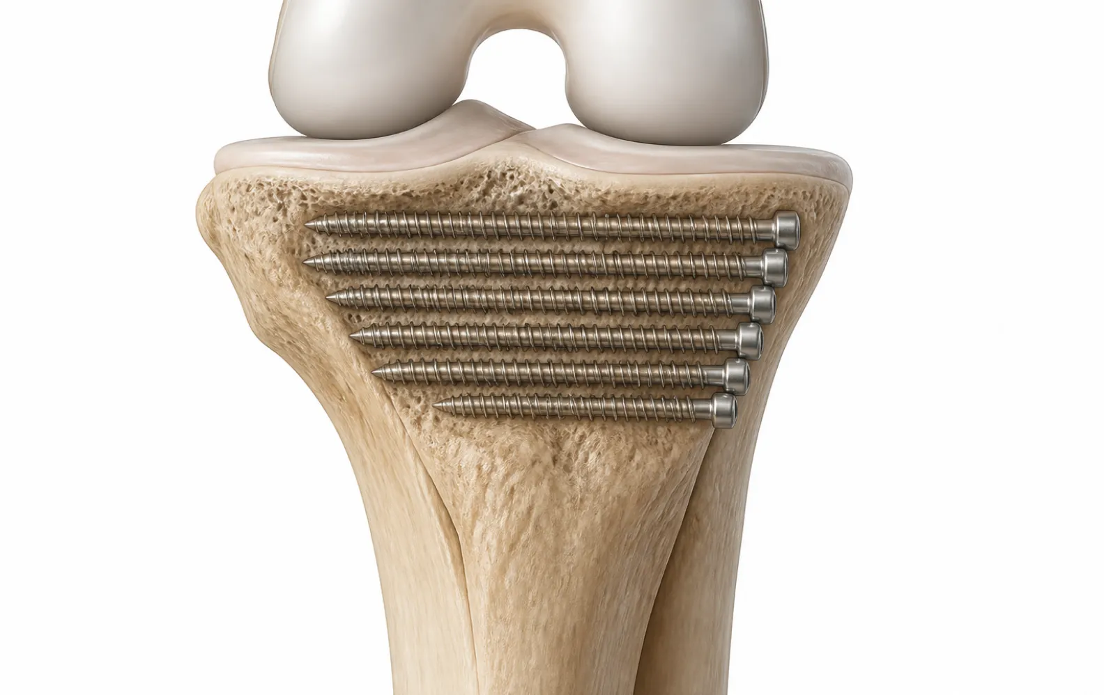
- Condylar Locking Screw Insertion: Calibrated 5.5mm cannulated locking screws are inserted over the guide wires into the condylar cluster. These screws advance through the plate head, purchasing dense subchondral bone across both condyles. In dense bone, self-tapping flutes evacuate bone debris, while double-lead threads ensure synchronized linear advance between the plate hole and bone matrix.
- Engaging the Oblique Strut Screw: In fractures with medial comminution, the oblique condylar strut screw is mandatory. Directed proximally and medially from the lower tier of the plate head, this 5.5mm screw crosses diagonally up to 70mm into the strong subchondral bone of the medial condyle. This creates a rigid internal triangular truss that resists severe varus bending loads. In fragile articular margins, smooth locking pegs can be alternated to prevent rotational torque on subchondral bone fragments.
Step 4: Diaphyseal Bridge Plating and Working Length Optimization
With the condylar block secured at 95° to the knee joint, attention turns to the femoral diaphysis. Diaphyseal fixation in comminuted fractures must adhere strictly to flexible bridge plating principles:
First, gross limb length, coronal alignment, and rotation are verified. Manual longitudinal traction or an articulated distractor spans the comminuted segment. Rotation is confirmed by assessing cortical thickness matching at the fracture boundaries and ensuring symmetrical hip internal/external rotation compared to the uninjured limb.
Second, if axial reduction requires drawing the plate flush to the bone, a non-locking cortical screw is placed through the dual compression slot in the plate shaft. Tightening this screw compresses the shaft against the bone, eliminating lateral standoff. Once anatomical reduction is achieved, bidirectional 4.5mm locking screws are inserted through the remaining diaphyseal holes. As detailed in our biomechanical study of locking plate versus conventional plate mechanics, leaving 3 to 4 empty plate holes over the comminuted fracture zone creates an optimal working length. This working length distributes bending strains evenly along the plate shaft, generating symmetrical 2% to 10% axial micromotion that promotes robust secondary callus formation.
Mechanical Assembly and Component Configuration
The following engineering matrix outlines standard component assembly for distal lateral femoral plating:
| Construct Zone | Hardware Selection | Diameter & Length Range | Mechanical Function |
|---|---|---|---|
| Metaphyseal Head Cluster | Cannulated Locking Screws | 5.5mm (30mm–100mm) | 95° fixed-angle parallel joint-line subchondral raft |
| Oblique Strut Position | Deep Medial Locking Screw | 5.5mm (Up to 70mm) | Triangulates condylar block; resists varus bending collapse |
| Shaft Dual Compression | Cortical Screws | 4.5mm (20mm–50mm) | Reduces bone to plate; provides optional interfragmentary compression |
| Shaft Bi-Cortical Locking | Self-Tapping Locking Screws | 4.5mm (26mm–55mm) | Extramedullary fixed-angle bridge anchorage; neutralizes shear |
Postoperative Rehabilitation and Construct Protection
Postoperative management must balance mechanical protection against joint mobilization:
- Immediate Range of Motion: Continuous passive motion (CPM) or active-assisted knee flexion is initiated within 24 to 48 hours postoperatively to prevent arthrofibrosis and intra-articular adhesions.
- Touchdown Weight-Bearing: In comminuted AO/OTA 33-C fractures, strict toe-touch or non-weight-bearing (10–15 kg) is maintained for 6 to 8 weeks. Axial loading during early recovery risks plate fatigue failure prior to bridging callus consolidation.
- Progression to Full Weight-Bearing: Progressive weight-bearing advances only after serial follow-up radiographs demonstrate bridging trabeculae across three of four cortices on orthogonal views, typically between weeks 10 and 14.
Formulary Management and Sourcing for Femoral Trauma
Maintaining institutional readiness for high-energy distal femoral trauma requires stocking complete plate-and-screw matrices. Because distal femoral fractures span diverse anatomies and comminution zones, surgical facilities must maintain 6, 10, 14, and 18-hole plate lengths in both left and right configurations. In complex multi-trauma presentations, alternative fixation protocols, such as a retrograde or antegrade cephalomedullary nail surgical technique, may be required depending on soft tissue envelopes and proximal fracture extension.
BoneCraft functions as a specialized global distributor of genuine orthopedic trauma implants. We maintain deep, ready-to-dispatch wholesale warehouse inventory covering complete femoral trauma implants and periarticular plating systems. Our products are supplied in factory-sealed sterile packaging with full lot traceability records, shielding healthcare systems from extended manufacturing lead times. Contact our technical team at Sale@bone-craft.com to discuss emergency hospital consignment or distributor volume arrangements.
Frequently Asked Questions
Why must articular reduction precede plate insertion in AO/OTA 33-C fractures?
Intra-articular fractures require anatomical reduction to prevent post-traumatic osteoarthritis. Because locking screws thread directly into the plate at pre-determined fixed angles, they cannot compress or manipulate displaced condylar fragments together. Articular congruity must be achieved first using independent lag screws before applying the bridging plate.
What is the mechanical advantage of the 95° fixed angle in the plate head?
The 95° angle reflects the anatomical relationship between the femoral mechanical axis and the distal femoral joint line. When the plate shaft is aligned flush with the lateral femoral diaphysis, the 95° condylar screw trajectory automatically aligns the subchondral screw raft parallel to the joint surface, restoring anatomical coronal alignment without manual plate bending.
How does the condylar strut screw prevent construct failure in comminuted fractures?
In fractures lacking medial cortical bone, axial weight-bearing exerts a severe varus bending moment on the lateral plate. The oblique strut screw drives proximally and medially across the condyles into the dense medial subchondral bone, creating an internal triangular truss. This truss converts cantilever bending forces into compressive vectors, reducing cyclic strain on the plate.
Why should plates not be over-filled with screws across the comminuted zone?
Placing screws directly into comminuted metaphyseal fragments devascularizes small bone chips and creates an overly rigid construct. Leaving 3 to 4 plate holes open over the fracture site increases the construct's working length, lowering peak bending stresses on the plate and promoting secondary bone healing through beneficial axial micromotion.
What is the role of the 102cm anatomical bow in distal femoral plating?
The human femoral shaft naturally bows anteriorly with a radius of curvature averaging roughly 100cm to 120cm. A plate engineered with a continuous 102cm anterior bow matches this natural curve, preventing the plate ends from impinging against the anterior cortex or pulling the femur into procurvatum malalignment.
When is a MIPO submuscular approach preferred over open exposure?
The MIPO submuscular technique is preferred in extra-articular fractures (AO/OTA 33-A) or intra-articular fractures with simple, easily reducible articular components. By inserting the plate through small incisions beneath the vastus lateralis, MIPO preserves periosteal blood flow and fracture hematoma, significantly accelerating biological bone healing and reducing infection risk.
-

BoneCraft Medical & Technical Editorial Team
Orthopedic Fixation Specialists
The BoneCraft Editorial Team comprises biomedical specialists and medical device supply chain experts. Dedicated to surgical transparency and technical precision, we publish insights on trauma plating, intramedullary fixation, and biomechanical implant performance. Our content synthesizes established surgical techniques, clinical literature, and exact specifications for genuine Zimmer Biomet orthopedic products to support global healthcare distributors and procurement teams.
