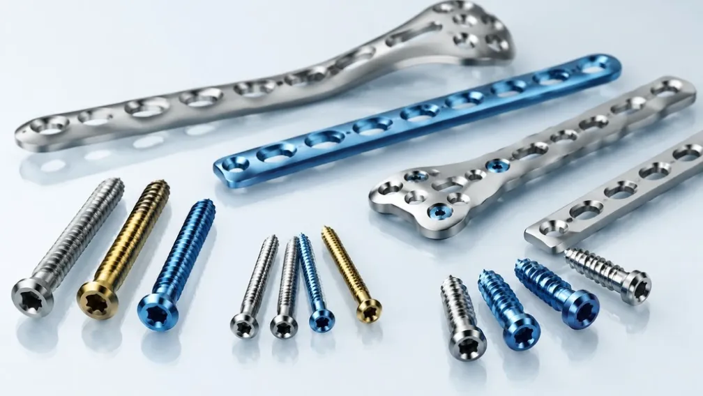
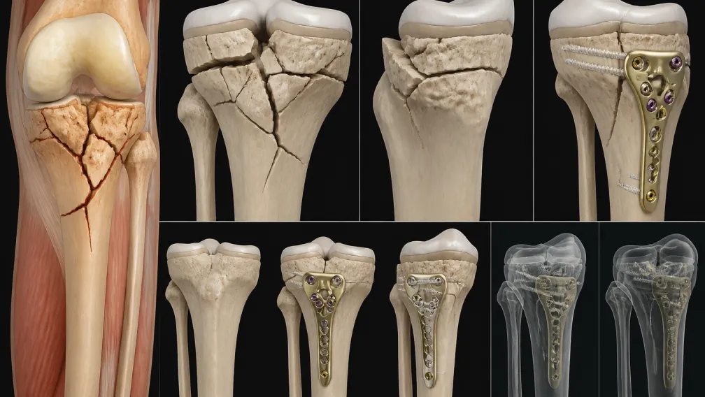
Intertrochanteric femur fractures are the most common hip fracture pattern treated operatively, with global incidence projected to rise substantially as the population over 65 expands. For trauma centers, ASCs, and implant distributors, the central implant decision in these cases comes down to two mature technologies: the extramedullary sliding hip screw (SHS) and the intramedullary cephalomedullary nail. Both achieve stable fixation and permit early weight bearing when applied to the correct fracture pattern. The clinical and economic differences between them emerge from fracture stability, lateral wall integrity, and the presence of subtrochanteric extension. This guide reviews the biomechanics, classification-driven indications, and clinical evidence behind each implant, then addresses the inventory implications for facilities stocking hip and femur trauma systems.
Fracture Classification and Stability Assessment
Implant selection begins with the AO/OTA 31-A classification, which stratifies trochanteric fractures by stability. Type A1 fractures are simple two-part patterns with an intact posteromedial buttress and lateral cortex; they are inherently stable once reduced. Type A2 fractures involve posteromedial comminution with loss of the calcar buttress, allowing uncontrolled collapse into varus and retroversion. Type A3 patterns, including transverse intertrochanteric and reverse obliquity fractures, lack any medial cortical contact and behave biomechanically like subtrochanteric fractures.
Lateral femoral wall integrity carries equal weight in this assessment. The lateral wall acts as a buttress that prevents excessive medial translation of the shaft during compression. Work by Hsu and colleagues demonstrated that a lateral wall thickness below 20.5 mm, measured on the anteroposterior view from a reference point 3 cm below the innominate tubercle, predicts secondary fracture displacement and fixation failure when a sliding hip screw is used. Thin lateral walls, reverse obliquity lines, and any extension into the subtrochanteric region shift the indication toward intramedullary fixation.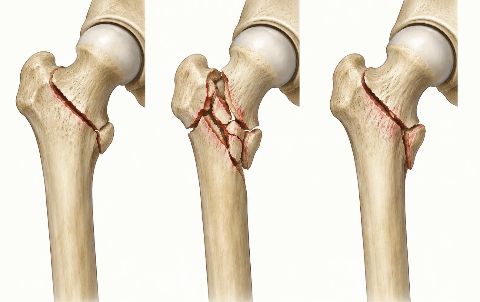
Sliding Hip Screw: Extramedullary Load Bearing
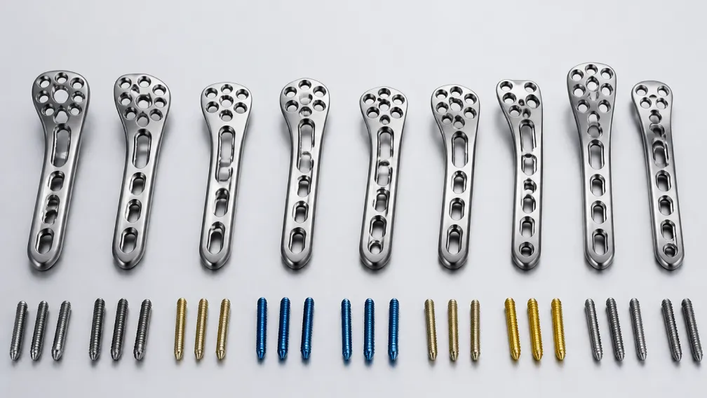
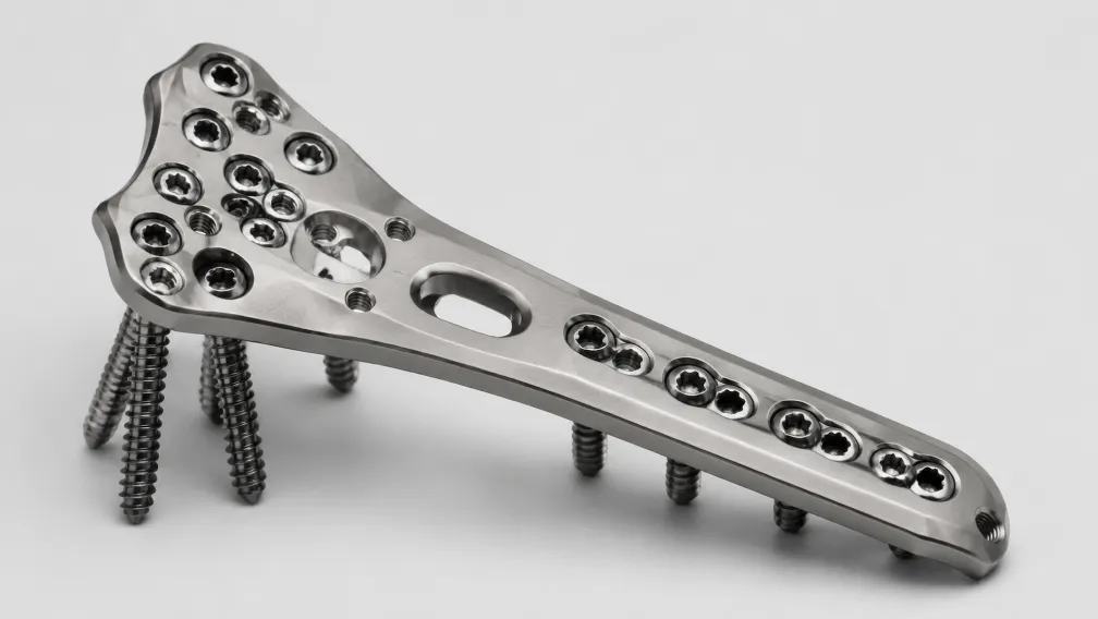
The sliding hip screw remains the reference implant for stable AO/OTA 31-A1 fractures. A large-diameter lag screw passes through the femoral neck into the head and slides within a barrel fixed to a side plate. During weight bearing, the fracture surfaces compress along the lag screw axis, converting shear forces into controlled impaction at the fracture site. This controlled collapse is a mechanical advantage in stable patterns because it increases bone-to-bone contact and stimulates union.
The implant carries practical advantages: low component cost, a short operative learning curve, and minimal reaming-related physiological insult. Its limitations appear in unstable patterns, where uncontrolled sliding produces limb shortening, shaft medialization, and abductor lever-arm dysfunction.
Tip-Apex Distance and Screw Position
Regardless of implant class, the position of the head element governs the risk of cut-out. Baumgaertner's tip-apex distance (TAD) quantifies this risk by summing the distance from the lag screw tip to the apex of the femoral head on anteroposterior and lateral views, corrected for magnification. A TAD below 25 mm is associated with a dramatic reduction in cut-out rates; values above that threshold should prompt intraoperative repositioning. The preferred screw trajectory is center-center or slightly inferior on the anteroposterior view and centered on the lateral view, seating the screw in dense subchondral bone.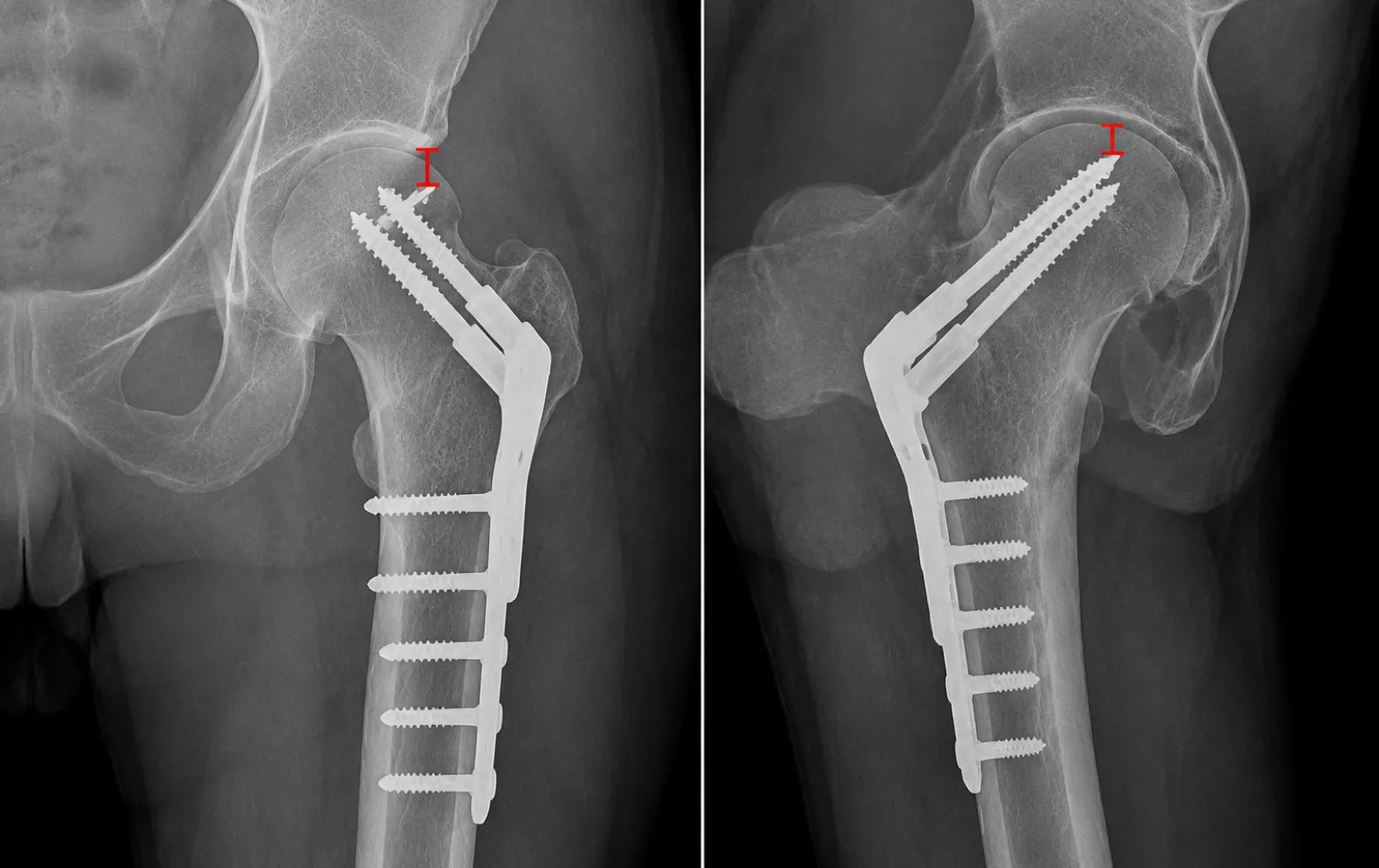
Cephalomedullary Nails: Intramedullary Load Sharing
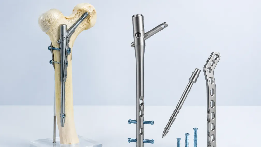
A cephalomedullary nail occupies the medullary canal and shares load along the mechanical axis of the femur. Compared with an extramedullary plate, the intramedullary position shortens the bending lever arm on the implant by roughly the distance between the lateral cortex and the canal center, reducing cantilever stress at the lag screw junction. The nail body also functions as an internal lateral wall buttress, capping the amount of shaft medialization and collapse in comminuted patterns.
Established indications include unstable A2 fractures with posteromedial comminution, all A3 and reverse obliquity patterns, fractures with subtrochanteric extension, pathological lesions, and cases where minimized limb shortening is a priority. Modern systems offer helical blades or lag screws as head elements; blade designs compact cancellous bone during insertion and show favorable cut-out behavior in osteopenic bone. Design differences between nail systems, including proximal geometry and locking options, are covered in our analysis of cephalomedullary nail designs.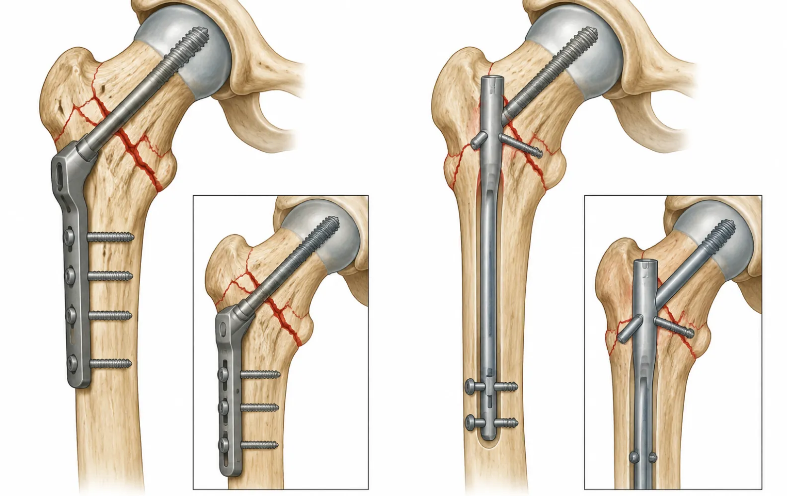
Short versus Long Nail Selection
A short cephalomedullary nail (typically 170 to 240 mm) suffices for standard intertrochanteric fractures with an intact diaphysis. It shortens operative time, reduces distal targeting steps, and lowers implant cost per case. A long cephalomedullary nail is indicated when the fracture extends into the subtrochanteric region, in metastatic or impending pathological fractures, and in atypical femoral fracture patterns associated with long-term bisphosphonate use. Long nails protect the entire femoral shaft but demand attention to anterior bow mismatch between the implant and the patient's femur, since distal anterior cortex perforation remains a documented complication in elderly patients with increased femoral bow.
What the Clinical Evidence Shows
Two decades of randomized trials, summarized in the Cochrane reviews by Parker and Handoll, support a pattern-specific conclusion. For stable trochanteric fractures, sliding hip screws and intramedullary nails produce equivalent mortality, functional recovery, and reoperation rates, while the sliding screw costs less and requires less operative time. For unstable patterns, particularly reverse obliquity and transverse intertrochanteric fractures, intramedullary nails reduce reoperation risk and preserve limb length and abductor mechanics.
Reported transfusion rates run slightly higher with nailing because of reaming and canal preparation, though modern unreamed or minimal-reaming insertion has narrowed this gap. Cut-out rates have fallen across both implant classes as TAD discipline has become standard practice. Current evidence gives no support for routine nailing of every trochanteric fracture; the economically and clinically rational position is a formulary that stocks both systems and assigns them by fracture pattern.
Health-economic modeling reinforces the pattern-based approach. In high-volume elderly trauma populations, the higher unit price of a nail is offset only when it prevents reoperations in unstable patterns; applied indiscriminately to stable fractures, routine nailing raises implant spend without measurable gains in mobility, residence status, or one-year mortality. Several national hip fracture audits and guideline bodies, including NICE, therefore continue to list the sliding hip screw as the default for stable trochanteric fractures and reserve intramedullary devices for unstable and reverse obliquity patterns.
Reduction Quality Governs Outcome
Implant choice cannot compensate for poor reduction. Varus malreduction and posterior sag of the shaft fragment are the two errors most strongly associated with failure in both systems. Closed reduction on a fracture table should restore neck-shaft angle and anteversion before any reaming or drilling begins; when closed manipulation fails, limited open reduction through the surgical approach is preferable to accepting a varus position. For nailing, the entry point at the tip or slightly medial to the tip of the greater trochanter determines proximal fragment alignment, since an entry point lateral to the canal axis drives the proximal fragment into varus during nail insertion. Detailed positioning, reduction, and insertion steps are outlined in our gamma nail surgical technique reference.
Inventory and Sourcing Implications for Hospitals and Distributors
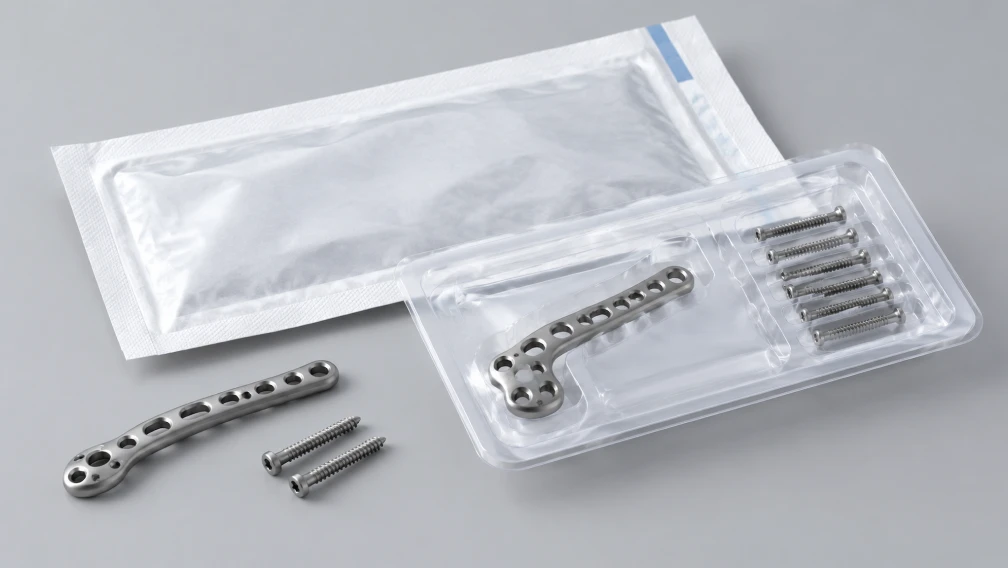
Procurement planning for a trochanteric fracture program should mirror the case mix. A general trauma hospital typically requires sliding hip screw sets for stable patterns alongside short and long nail options for unstable and extended fractures, with helical blade and lag screw head elements available in the diameters and lengths matched to the local patient population. Instrument compatibility, sterile versus non-sterile packaging, sterilization lead time, and consignment arrangements all affect total cost of ownership more than unit implant price.
Distributors building a trauma portfolio benefit from consolidating these items with a manufacturer that covers the full trochanteric spectrum, including nails, side plates, and matched screws, under one quality system and one logistics channel. BoneCraft supplies this portfolio through its orthopedic implant wholesale program, with batch traceability and documentation aligned to hospital tender requirements.
Summary
The decision algorithm is stable across the literature. Stable AO/OTA 31-A1 fractures with an intact lateral wall: sliding hip screw, TAD under 25 mm. Unstable A2 patterns, reverse obliquity or A3 fractures, subtrochanteric extension, and pathological bone: cephalomedullary nail, with length matched to the extent of disease. Both implants fail predictably when reduction is poor or the head element sits high in the femoral head, so technique discipline matters as much as implant selection. Facilities that stock both systems and apply them by pattern achieve the best combination of clinical outcome and implant spend.
For product specifications, available sizes, and distributor terms across our hip and femur trauma line, contact the BoneCraft team for a current catalog and quotation.
-

BoneCraft Medical & Technical Editorial Team
Orthopedic Fixation Specialists
The BoneCraft Editorial Team comprises biomedical specialists and medical device supply chain experts. Dedicated to surgical transparency and technical precision, we publish insights on trauma plating, intramedullary fixation, and biomechanical implant performance. Our content synthesizes established surgical techniques, clinical literature, and exact specifications for genuine Zimmer Biomet orthopedic products to support global healthcare distributors and procurement teams.