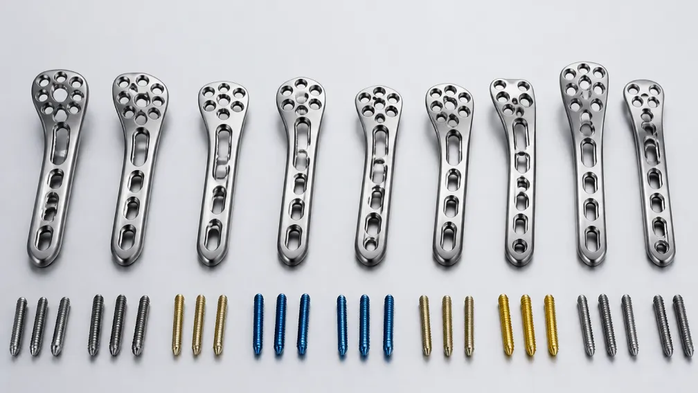
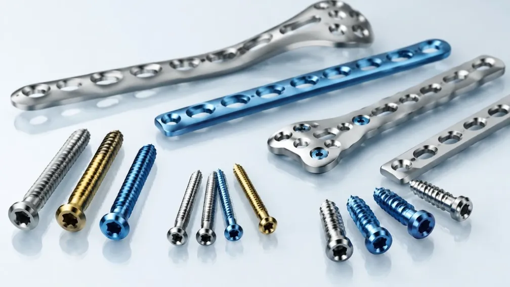
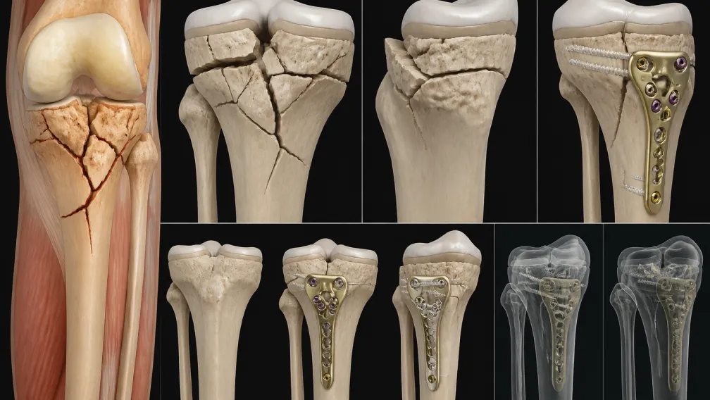
Proximal humerus fractures account for approximately 5% to 6% of all adult fractures, representing the third most common fracture pattern in patients over the age of 65. Driven by the increasing prevalence of osteoporosis, low-energy falls frequently result in complex 3-part and 4-part fracture configurations according to the Neer classification. While non-operative management suffices for minimally displaced fractures, unstable multi-fragmentary injuries demand rigid surgical stabilization to restore shoulder kinematics. Historically, operative fixation with conventional non-locking plates yielded unacceptably high failure rates due to screw toggle in cancellous bone and subsequent varus collapse. The introduction of anatomically contoured fixed-angle locking plates revolutionized shoulder and humerus trauma management. However, mechanical success relies entirely on adhering to strict biomechanical principles—specifically, the restoration of medial calcar support and precise multi-planar screw configuration. This analysis deconstructs the engineering mechanics of proximal humerus plating and the supply chain requirements for trauma formularies.
The Biomechanical Challenge: Deforming Forces and Varus Collapse
The proximal humerus is subjected to massive, multi-directional deforming forces exerted by the rotator cuff musculature. In a displaced 3-part or 4-part fracture, the supraspinatus and infraspinatus pull the greater tuberosity superiorly and posteriorly. The subscapularis pulls the lesser tuberosity medially. Simultaneously, the pectoralis major and deltoid displace the humeral shaft medially and proximally. The net mechanical vector of these unopposed forces drives the articular humeral head fragment into severe varus angulation.
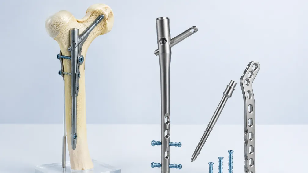
When a surgeon reduces the fracture and applies a lateral plate, the implant must act as a load-bearing cantilever beam. If the medial cortical hinge (the medial calcar) is comminuted or lacks bony contact, the entire physiological bending moment is transferred directly to the plate-bone interface. In osteoporotic bone, conventional screws rely on friction and thread purchase. Under cyclic loading, these screws simply cut out through the soft cancellous bone of the humeral head, resulting in catastrophic varus collapse, loss of reduction, and intra-articular screw penetration. Transitioning to a fixed-angle construct fundamentally alters this failure mechanism, as detailed in the structural principles of locking plate biomechanics.
The Imperative of Medial Calcar Support
The single most critical determinant of construct survival in proximal humerus plating is the establishment of medial calcar support. The medial calcar is the dense cortical bone at the inferomedial neck of the humerus. A proximal humerus locking plate must not merely act as a lateral tension band; it must physically prop up the medial column to neutralize varus bending forces.
Modern anatomical locking plates achieve this via dedicated inferomedial locking screw trajectories—commonly referred to as "calcar screws." These holes are located in the distal tier of the plate head and are angled obliquely upward. When inserted, the calcar screws drive tangentially against the medial cortical arch of the humeral head, acting as a rigid structural "kickstand." Clinical biomechanical studies demonstrate that placing at least two divergent inferomedial calcar screws increases the axial load-to-failure limit by over 40%, effectively shielding the superior screws from cyclic cantilever fatigue and preventing the humeral head from subsiding into varus.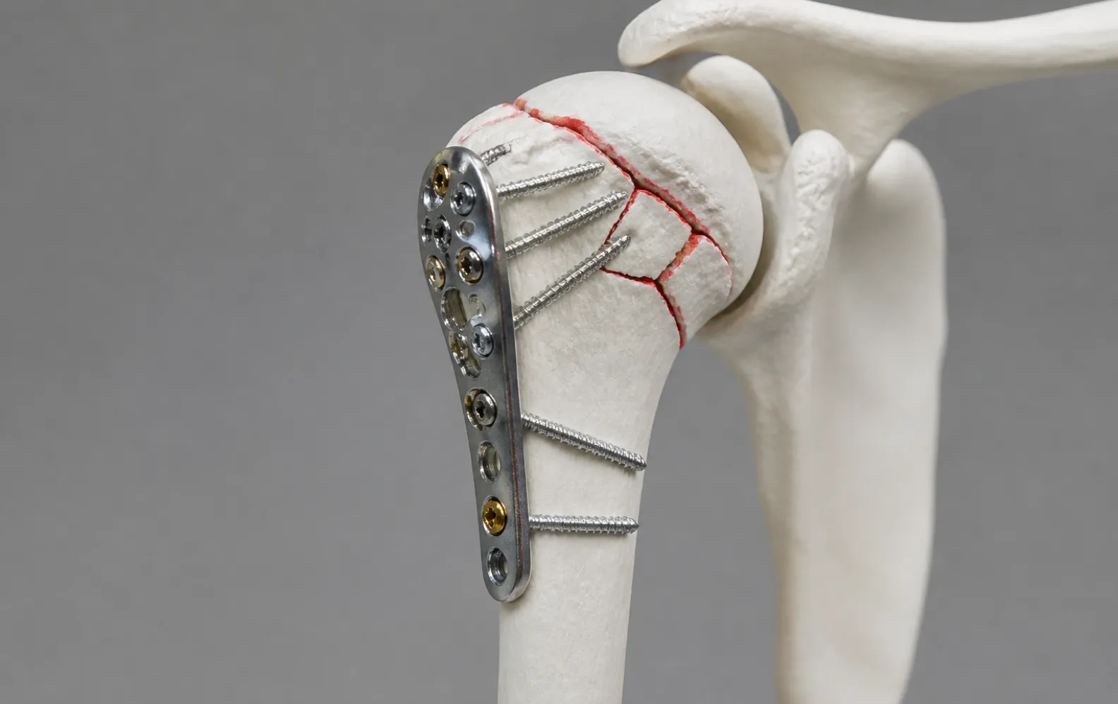
Multi-Planar Screw Configurations: Pegs vs. Threaded Screws
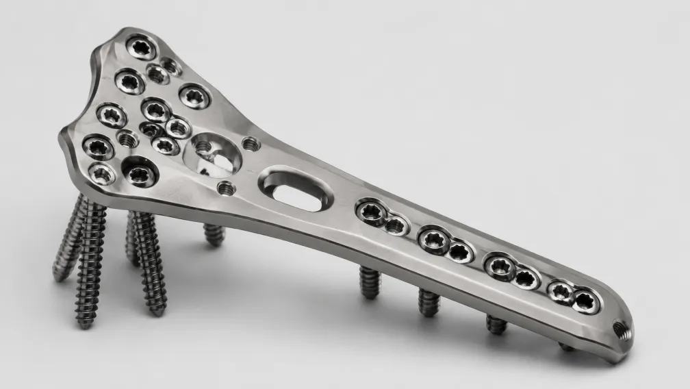
The humeral head consists of a thin subchondral shell encasing a low-density cancellous core. To maximize fixation strength, the proximal head of the locking plate features multiple divergent and convergent threaded holes. This multi-planar array creates a three-dimensional scaffold that captures fragments across the anterior, posterior, and superior quadrants of the humeral head.
Surgeons must deliberately choose between fully threaded locking screws and smooth locking pegs for this proximal cluster. Threaded locking screws provide maximum pull-out resistance, making them ideal for capturing large, dense bone fragments. However, in severe osteopenia, if microscopic varus collapse does occur during the healing phase, the sharp threads of standard screws will aggressively lacerate the glenoid cartilage, causing irreversible post-traumatic osteoarthritis.
To mitigate this risk, many advanced systems deploy proximal humerus plate locking pegs. These implants feature a threaded head that locks rigidly into the plate, but possess a completely smooth, unthreaded cylindrical shaft. The blunt-tipped pegs act as subchondral support beams. They provide identical resistance to varus bending moments, but if marginal collapse occurs, the blunt, threadless tips are significantly less destructive to the opposing glenoid articular surface. Furthermore, smooth pegs eliminate bony ingrowth along the shaft, facilitating atraumatic removal during future elective hardware extraction or conversion to arthroplasty.
Soft Tissue Management and Tuberosity Fixation
Beyond osseous fixation, functional recovery depends heavily on rotator cuff integrity and preserving the precarious blood supply to the humeral head. The primary arterial feed to the articular segment is the ascending branch of the anterior circumflex humeral artery, which runs in the bicipital groove. Overzealous surgical dissection, extensive periosteal stripping, or placing the plate too far anteriorly disrupts this vascular supply, leading to avascular necrosis (AVN) of the humeral head.
Proper plate positioning is equally vital for avoiding subacromial impingement. The superior edge of the plate must be positioned at least 5mm to 8mm distal to the tip of the greater tuberosity. If placed too proximally, the rigid titanium plate will impinge against the acromion during shoulder abduction, causing intractable pain and rotator cuff attrition.
Furthermore, locking screws alone are often insufficient to hold osteoporotic, fragmented tuberosities against the pull of the rotator cuff. Therefore, premium proximal humerus plates integrate dedicated suture holes along the proximal margins. Surgeons pass heavy, non-absorbable sutures through the rotator cuff tendon insertions and tie them directly into the plate. This tension-band suturing neutralizes the displacing muscle forces, compresses the tuberosities against the head-shaft junction, and offloads mechanical stress from the locking screws.
Material Science: Titanium Alloy versus Stainless Steel
Proximal humerus plates are manufactured in both 316L (or 22-13-5) stainless steel and Ti-6Al-4V titanium alloy, each presenting distinct mechanical trade-offs.
| Biomechanical Parameter | Ti-6Al-4V Titanium Alloy | 316L / 22-13-5 Stainless Steel |
|---|---|---|
| Elastic Modulus (Stiffness) | ~110 GPa (Closer to cortical bone) | ~200 GPa (Highly rigid) |
| Stress Shielding Risk | Low; allows beneficial micromotion for callus formation | Higher; absorbs load, risking osteopenia beneath plate |
| Intraoperative Contouring | Resists manual bending; notch-sensitive | Easier to contour to atypical anatomies without failure |
| MRI Artifact | Minimal distortion for postoperative imaging | Significant artifact scattering |
For the proximal humerus, titanium is predominantly favored. Its lower modulus of elasticity reduces construct stiffness, mitigating the risk of the hard plate cutting through osteopenic bone at the proximal screw interface. Additionally, because postoperative assessment of rotator cuff integrity and AVN progression often requires MRI, the minimal magnetic artifact of titanium provides a critical diagnostic advantage.
Hospital Procurement and Inventory Sourcing Logistics
Managing inventory for upper extremity trauma presents distinct challenges for central sterile supply departments (CSSDs) and regional medical device distributors. Proximal humerus fractures are acute emergencies, yet the anatomical variability among patients necessitates a dense matrix of implant configurations. Trauma centers must maintain left- and right-specific anatomical plates in lengths spanning 3-hole (90mm) to 12-hole (240mm) variants to address everything from isolated surgical neck fractures to extensive diaphyseal comminution.
Stockouts of calcar screws, smooth locking pegs, or specific plate lengths compromise surgical outcomes and force delays. As global contract manufacturing bottlenecks extend lead times across major OEMs, hospitals face increasing vulnerability. Bridging this supply chain gap requires partnering with dedicated independent stockists capable of immediate fulfillment.
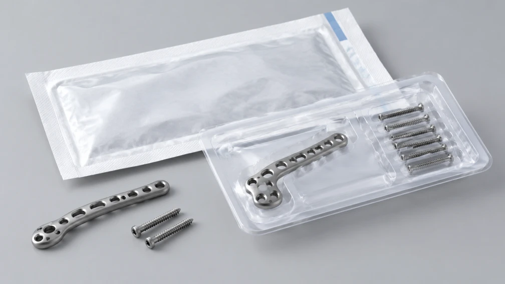
BoneCraft functions as a specialized independent distributor of genuine, verified orthopedic trauma implants. We decouple hospital procurement from OEM manufacturing delays by maintaining deep, accessible wholesale warehouse inventory. Our sterile-packaged trauma portfolios include complete left/right proximal humerus plating sets, modular locking screw matrices, and advanced instrumentation. Every component is shipped with complete lot-level traceability and regulatory compliance documentation, ensuring your trauma suites remain fully operational for complex extremity reconstructions. Contact our distribution team to review our current stock availability and consignment terms.
Frequently Asked Questions
Why is medial calcar support critical in proximal humerus fracture fixation?
The medial calcar serves as the anatomical hinge that resists the massive varus bending forces exerted by the rotator cuff and deltoid muscles. If the medial cortex is comminuted or lacks bony contact, the locking plate bears the entire physiological load as a cantilever. Inserting inferomedial locking screws (calcar screws) provides a rigid structural kickstand against the medial arch, dramatically increasing the construct's resistance to varus collapse and screw cut-out.
How do surgeons prevent subacromial impingement during plate placement?
Subacromial impingement occurs when the locking plate is positioned too high on the humerus, causing the rigid metal to strike the acromion during shoulder abduction. To prevent this, surgeons must position the superior edge of the plate at least 5mm to 8mm distal to the proximal tip of the greater tuberosity, confirming the clearance dynamically under intraoperative fluoroscopy before final screw fixation.
What is the primary cause of screw cut-out in the humeral head?
Screw cut-out typically results from a combination of severe osteoporosis, a lack of medial calcar support, and rigid plate constructs. When bending moments overload the fixation, the hard titanium screws slice upward through the soft, low-density cancellous bone of the humeral head, ultimately penetrating the articular cartilage and damaging the glenoid.
When are smooth locking pegs preferred over fully threaded screws?
Smooth locking pegs are deployed in the proximal subchondral clusters in patients with severe osteopenia. Because they lack threads on the shaft, they provide excellent blunt cantilever support against subsidence. If marginal varus collapse does occur, the blunt tips are significantly less destructive to the opposing glenoid cartilage compared to the sharp threads of conventional locking screws.
Why do modern proximal humerus plates feature suture holes?
Locking screws alone frequently fail to secure fragmented, osteoporotic tuberosities against the powerful pull of the supraspinatus and subscapularis muscles. The suture holes allow surgeons to pass heavy, non-absorbable sutures through the rotator cuff tendon insertions, tying the tuberosities down directly to the plate. This tension-band effect neutralizes displacing muscle forces and offloads mechanical stress from the primary bone screws.
Why is titanium favored over stainless steel for proximal humerus plating?
Titanium (Ti-6Al-4V) possesses a lower elastic modulus than stainless steel, making the plate less rigid. This reduced stiffness shares load more evenly with the osteopenic bone, reducing the risk of screw cut-out. Additionally, titanium produces significantly less magnetic artifact on postoperative MRI, which is essential for monitoring rotator cuff healing and diagnosing avascular necrosis (AVN) of the humeral head.
-

BoneCraft Medical & Technical Editorial Team
Orthopedic Fixation Specialists
The BoneCraft Editorial Team comprises biomedical specialists and medical device supply chain experts. Dedicated to surgical transparency and technical precision, we publish insights on trauma plating, intramedullary fixation, and biomechanical implant performance. Our content synthesizes established surgical techniques, clinical literature, and exact specifications for genuine Zimmer Biomet orthopedic products to support global healthcare distributors and procurement teams.