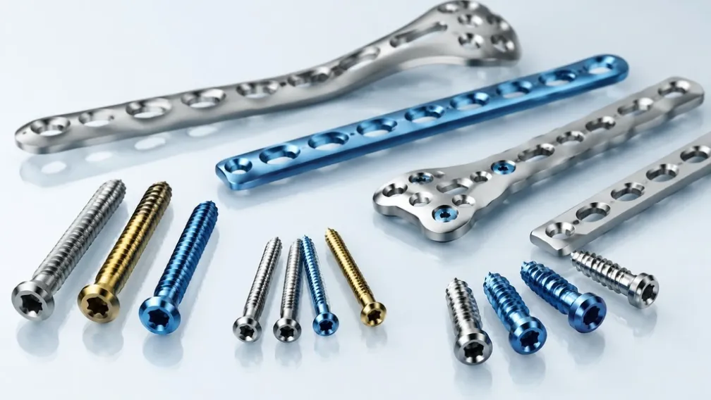
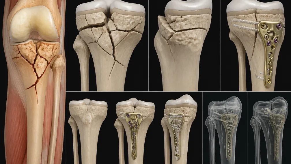
Fractures of the distal radius represent the most frequently encountered skeletal injury in adult trauma care, accounting for nearly one-sixth of all fractures evaluated in emergency departments and a predominant share of operative wrist and hand fractures. The demographic distribution is bimodal: high-energy motor vehicle and athletic trauma in young adults, contrasted with low-energy insufficiency falls on an outstretched hand in elderly patients with osteopenic bone. Historically, closed reduction and cast immobilization or percutaneous pinning struggled to maintain anatomical alignment in unstable, intra-articular fractures. The advent of dorsal plating resolved dorsal comminution but introduced significant soft tissue morbidity, including extensor pollicis longus (EPL) and extensor digitorum communis (EDC) tenosynovitis or rupture due to the subcutaneous prominence of dorsal implants. The clinical transition to fixed-angle volar locked plating revolutionized distal radius management by capitalizing on the spacious volar pronator fossa, establishing an extramedullary subchondral scaffold that neutralizes articular displacement while shielding surrounding tendons.
Biomechanical Foundation: The Three-Column Theory of the Wrist
Modern surgical fixation of intra-articular distal radius fractures relies on the three-column concept originally formalized by Rikli and Regazzoni. The wrist is divided into three distinct load-bearing longitudinal columns across the distal forearm, each carrying specific biomechanical duties:
- The Radial Column (Lateral): Encompasses the radial styloid and the scaphoid fossa. It serves as an anatomical strut providing osseous attachment for the robust brachioradialis tendon and the extrinsic radiocarpal ligaments. It resists radial deviation and tensile distraction under power grip.
- The Intermediate Column (Central): Encompasses the lunate fossa and the sigmoid notch of the radius. This column is the primary weight-bearing pillar of the human wrist, transmitting approximately 60% to 80% of axial compressive joint reactive forces directly across the radiocarpal articulation. Restoration of the lunate facet is critical; residual step-offs exceeding 1mm to 2mm exponentially accelerate radiocarpal arthritis.
- The Ulnar Column (Medial): Encompasses the distal ulna, the triangular fibrocartilage complex (TFCC), and the distal radioulnar joint (DRUJ). It stabilizes forearm pronation-supination rotation and balances transverse shear loads across the wrist.
In complex AO/OTA 23-C2 and C3 intra-articular fractures, comminution frequently splits the intermediate column into dorsal and volar rim fragments (die-punch fragments), alongside a sheared radial styloid. Restoring functional kinematics requires internal fixation hardware capable of providing rigid subchondral neutralization across both the radial and intermediate columns simultaneously.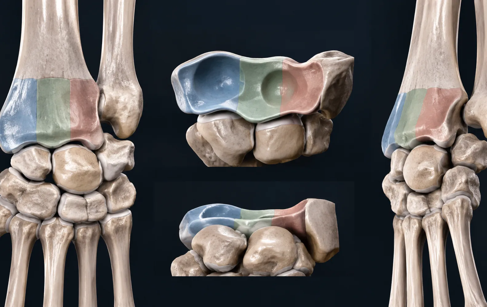
Volar Anatomy and the Critical Watershed Line
The primary advantage of the volar approach is the protective soft-tissue envelope provided by the pronator quadratus muscle and the deep flexor compartment. However, achieving stability without inciting flexor tendon attrition demands rigorous anatomical discipline regarding the "watershed line."
The watershed line is defined as the theoretical transverse margin marking the most distal edge of the pronator quadratus insertion, located approximately 2mm proximal to the radiocarpal joint capsule at the level of the scaphoid fossa and within millimeters of the joint margin at the lunate fossa. Distal to this ridge, the volar cortex sweeps dorsally, creating an intimate relationship with the flexor tendons—most notably the flexor pollicis longus (FPL) and the flexor digitorum profundus (FDP) to the index finger.
When an implant is positioned distal to the watershed line or sits proud off the volar cortex, repetitive excursion of the FPL tendon over the hardware margin induces chronic friction, fraying, and eventual attritional rupture. To prevent this devastating complication, modern pre-contoured plates such as the DVR volar distal radius plate and the anatomic distal radius plate incorporate low-profile chamfered distal edges, specific sub-watershed footprints, and a built-in 11° to 12° volar tilt that mimics native anatomy, minimizing hardware impingement.
Subchondral Raft Mechanics and Variable Screw Vectors
Because the distal radius features thin cortical bone and porous metaphyseal trabeculae, conventional non-locked screws frequently strip or toggle under cyclic physiological loading. Fixed-angle volar locking plates overcome this by functioning as an internal fixator, where screws thread directly into the plate head.
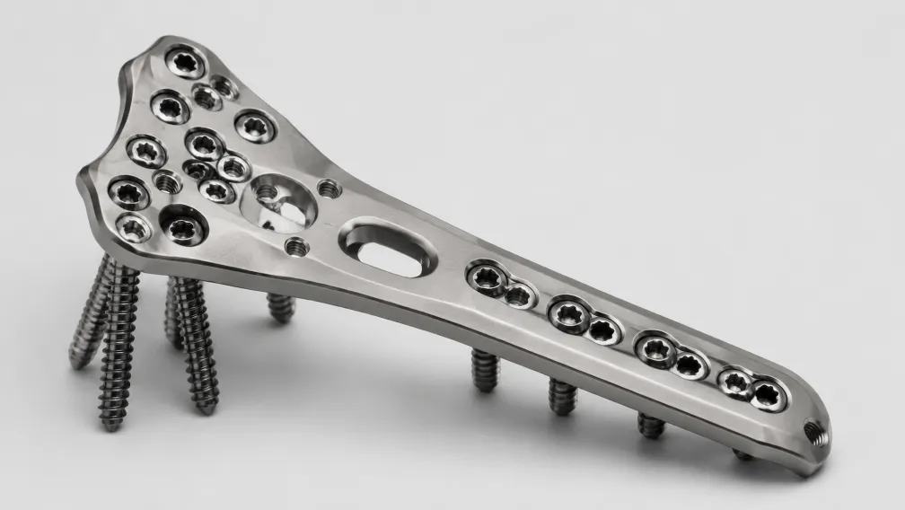
The distal head of the plate incorporates multi-tiered, divergent locking screw trajectories engineered to form a subchondral "raft." In advanced plating constructs, the distal-most row of locking holes directs screws immediately beneath the articular cartilage of the scaphoid and lunate fossae. These screws run within 2mm to 4mm of the articular margin, creating a horizontal titanium or stainless steel shelf that physically blocks depressed osteochondral fragments from sinking proximally into the metaphyseal void.
In severely comminuted or osteoporotic articular blocks, surgeons frequently deploy smooth locking pegs in place of fully threaded screws in the distal subchondral cluster. Smooth pegs provide identical cantilever beam rigidity against axial joint depression while eliminating rotational torsion during insertion and avoiding osseous integration along the shaft, which ensures atraumatic hardware removal during future revision procedures.
Furthermore, when high-energy impact shears off a separate radial styloid fragment, standard central plate screws may fail to capture the lateral cortex. In such fracture configurations, supplemental lateral column stabilization using a dedicated low-profile radial styloid plate or modular buttress pin provides isolated, rigid fixation that resists brachioradialis distraction forces.
Mechanical Performance: Volar Locked Plating vs. Historical Constructs
The superiority of pre-contoured volar locking technology over historical fixation options is evident across key mechanical and biological parameters:
| Construct Metric | Closed Reduction & Casting | Dorsal Non-Locked Plating | Pre-Contoured Volar Locked Plate |
|---|---|---|---|
| Biomechanical Stability | Low; subject to early cast loosening | Moderate; relies on cortical friction | Superior; fixed-angle subchondral beam scaffold |
| Soft Tissue Envelope Coverage | High swelling / cast pressure risk | Poor; thin dorsal subcutaneous layer | Excellent; protected beneath pronator quadratus |
| Tendon Complication Profile | Secondary tendon stiffness | High EPL / EDC extensor tenosynovitis & rupture | Low FPL risk when placed proximal to watershed line |
| Maintenance of Volar Tilt | Frequent secondary dorsal collapse (>30%) | Moderate maintenance; plate fatigue risk | Predictable; fixed-angle screws resist dorsal moment |
| Postoperative Mobilization | Delayed (6–8 weeks immobilization) | Moderate (protected 3–4 weeks) | Immediate active-assisted motion within 1–2 weeks |
Surgical Execution: Henry Approach and Component Sequencing
Achieving consistent outcomes with volar locked plating mandates systematic procedural sequencing. The primary steps executed during open reduction internal fixation (ORIF) include:
- Surgical Approach: The modified flexor carpi radialis (FCR) approach or classical Henry approach is performed. A longitudinal incision is placed over the FCR tendon sheath. The sheath is incised, and the tendon is retracted ulnarly, protecting the median nerve within the carpal tunnel. The deep fascia is incised to expose the pronator quadratus.
- Pronator Quadratus Elevation: An L-shaped incision is made through the radial and distal margins of the pronator quadratus, leaving a small cuff of tissue on the radius for later repair. The muscle belly is carefully elevated subperiosteally to expose the volar radius while preserving the joint capsule.
- Articular Reduction: The fracture hematoma is cleared. Under traction and manual manipulation, radial length, radial inclination (normal 22°–23°), and volar tilt (normal 11°–12°) are provisionally restored. Temporary fixation is held with 1.6mm Kirschner wires directed subchondrally or from the radial styloid.
- Plate Positioning & Diaphyseal Reference: The volar locking plate is positioned on the volar cortex, taking care to seat the distal margin precisely proximal to the watershed line. A non-locking cortical screw is initially inserted through the central elongated gliding slot in the plate shaft. This oblong hole allows fine-tuning of longitudinal plate position under fluoroscopy before final distal locking.
- Distal Subchondral Locking: Threaded drill guides are attached to the distal plate head. Calibrated 2.4mm or 2.7mm locking screws are drilled and measured. Screws must engage subchondral bone within 2mm of the articular margin without penetrating the radiocarpal joint or breaching the dorsal cortex, where overlong screws can fray extensor tendons in the third or fourth dorsal compartments. Detailed step-by-step techniques are outlined in our reference guide on volar locking plate distal radius technique execution.
- Diaphyseal Locking and Closure: The remaining shaft holes are filled with 3.5mm cortical or locking screws according to locking plate biomechanics principles. The pronator quadratus is re-approximated over the plate using absorbable sutures to provide a biological barrier between the hardware and the overlying flexor tendons.
Soong Grading and Flexor Tendon Protection
A comprehensive clinical evaluation of volar plating requires assessing the relationship between implant prominence and the volar cortical rim, standardized by Soong et al.:
- Soong Grade 0: The plate sits entirely proximal to the watershed line and remains dorsal to a line drawn tangential to the volar rim. The risk of FPL tendon irritation is minimal (<1%).
- Soong Grade 1: The plate is positioned at or slightly distal to the watershed line but remains dorsal to the volar rim projection. Close postoperative monitoring is recommended.
- Soong Grade 2: The plate extends distal to the watershed line and sits proud, protruding volar to the tangential line of the volar cortical margin. Grade 2 placement dramatically escalates the risk of catastrophic FPL tendon attrition and represents a strong indication for elective hardware removal once bone healing is confirmed.
Formulary Management and Sourcing for High-Volume Trauma
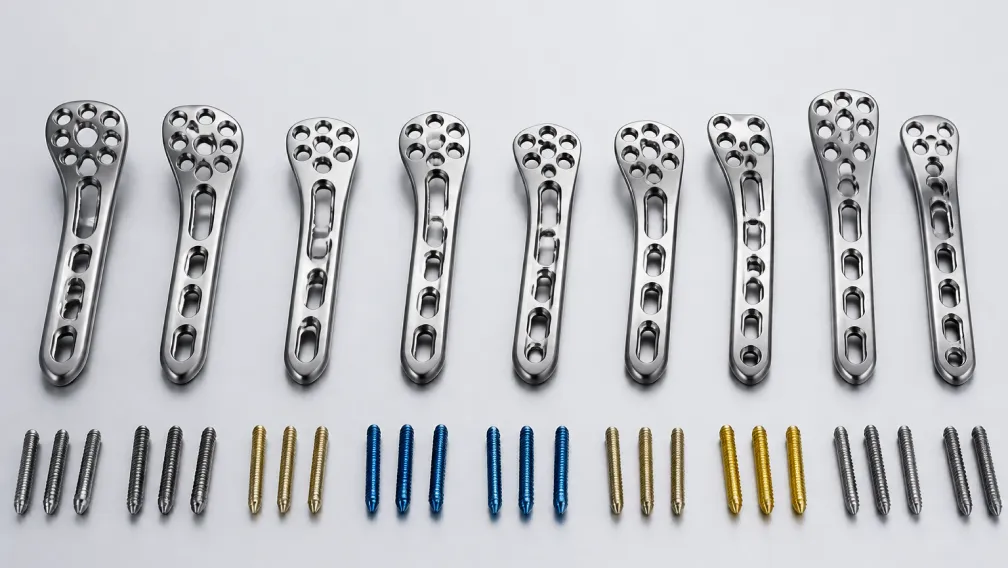
Because distal radius fractures occur constantly in both ambulatory surgery centers (ASCs) and acute hospital trauma centers, managing wrist and hand fixation inventory demands immediate hardware availability across a dense matrix of anatomical profiles:
Formularies cannot rely on a single plate geometry. Modern trauma centers must stock standard, narrow, and wide head widths across both left and right configurations, alongside 2.4mm, 2.7mm, and 3.5mm screw lengths spanning 10mm to 30mm. When contract device manufacturers face global supply constraints or backorders, surgical suites face case cancellations or forced compromises in plate width that jeopardize lateral or intermediate column capture.
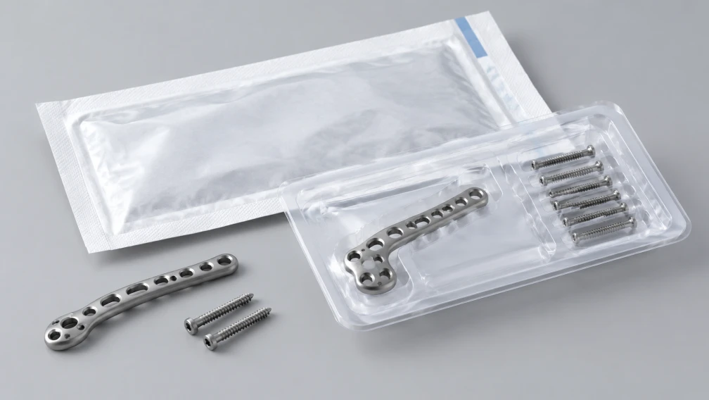
BoneCraft resolves these supply chain vulnerabilities by functioning as a dedicated independent distributor of genuine trauma systems. We maintain robust, ready-to-ship wholesale warehouse inventory covering authentic manufacturer-packaged volar plating trays, modular cannulated systems, and anatomical extremity sets. All products carry intact lot numbers, full trace documentation, and verified regulatory certifications. Contact our surgical distribution team at Sale@bone-craft.com to review live warehouse availability and hospital consignment agreements.
Frequently Asked Questions
What is the watershed line, and why is it critical in volar locking plate placement?
The watershed line is the transverse anatomical ridge marking the distal limit of the pronator quadratus muscle and the attachment of the volar radiocarpal joint capsule. Placing a plate distal to this ridge causes the implant to sit proud over the volar rim, bringing sharp metal corners into direct contact with the flexor pollicis longus (FPL) tendon. Chronic friction during repetitive finger and thumb flexion can lead to FPL tendon fraying and attritional rupture.
How does the three-column theory dictate internal fixation in intra-articular fractures?
The three-column theory separates the distal radius into the radial column (radial styloid), intermediate column (lunate fossa and sigmoid notch), and ulnar column (distal ulna and DRUJ). Because the intermediate column transmits over 60% of axial load across the wrist, failure to reduce and support the lunate fossa leads to joint incongruity, dorsal tilt, and post-traumatic arthritis. Volar locking plates must feature distal screw clusters that cross under both the scaphoid and lunate facets to restore dual-column stability.
Why are smooth locking pegs used instead of threaded locking screws in subchondral bone?
Smooth locking pegs provide fixed-angle cantilever support directly beneath the articular surface without cutting into brittle subchondral fragments during insertion. Unlike threaded screws, smooth pegs do not rotate or shear fragile cartilage fragments during tightening. Furthermore, bone does not grow into thread flutes, dramatically reducing torque requirements and preventing bone destruction during subsequent hardware removal.
What radiographic parameters define an acceptable reduction of the distal radius?
Standard radiographic criteria for an acceptable reduction include: radial inclination of 15° to 25° (normal ~22°), volar tilt between neutral (0°) and 15° volar tilt (normal ~11°–12°), radial length discrepancy within 2mm to 3mm of the uninjured contralateral wrist, and intra-articular step-off or gap less than 1mm to 2mm on orthogonal fluoroscopic views.
How does the Soong grading system assess the risk of flexor tendon rupture?
The Soong grading system evaluates plate prominence relative to the volar cortical rim on a true lateral radiograph. Grade 0 indicates the plate sits entirely proximal to the watershed line (low risk). Grade 1 indicates the plate is close to the watershed line but does not project volar to the rim. Grade 2 indicates the plate sits distal to the watershed line and projects prominently volar to the anterior cortex, representing a high risk for FPL tendon rupture that often warrants elective hardware removal.
Why must ASCs and trauma hospitals maintain inventory across multiple plate widths?
Patient anatomy varies widely in transverse distal radius dimensions. A standard plate placed on a narrow wrist overhangs the radial or ulnar borders, causing soft tissue irritation. Conversely, a narrow plate placed on a broad wrist fails to capture the radial styloid or the volar-ulnar lunate facet. Facilities must maintain narrow, standard, and wide plate variations to ensure complete column capture across all patient sizes.
-

BoneCraft Medical & Technical Editorial Team
Orthopedic Fixation Specialists
The BoneCraft Editorial Team comprises biomedical specialists and medical device supply chain experts. Dedicated to surgical transparency and technical precision, we publish insights on trauma plating, intramedullary fixation, and biomechanical implant performance. Our content synthesizes established surgical techniques, clinical literature, and exact specifications for genuine Zimmer Biomet orthopedic products to support global healthcare distributors and procurement teams.